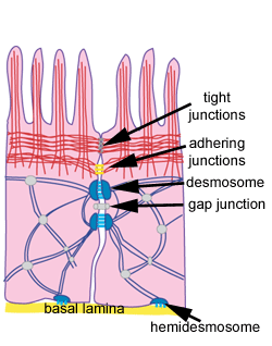
Junctions
Adhering Junctions
Epithelial cells are held together by strong anchoring (adherens)
junctions.
There are two types of adherens junctions:
zonula adherens - which contain actin filaments
macula adherens (desmosomes) which contain intermediate filaments.
The zonula adherens junction lies below the tight junction (occluding
junction). In the gap between the two cells, there is a protein
called E-cadherin - a cell membrane glycoprotein. The cadherins from
adjacent cells interact to 'zipper' up the two cells together.
Inside the cell, E-cadherin binds to catenin, which in turn binds to other proteins (vinculin, alpha actinin) in a protein complex with actin filaments (microfilaments, shown here in
red).
The actin filaments tend to
be arranged circumferentially around the cell, into what is called a 'marginal' band. This marginal band can contract, and deform the shape of cells
held together in this way - this is thought to be key in the morphogenesis
of epithelial cells, in forming ducts for example.
Tight junctions
These are regions in which two cells are very tightly connected
together, and they will prevent some molecules from passing across
an epithelium. The borders of two cells are fused together, often
around the whole perimeter of each cell, forming a continuous belt
like junction known as a tight junction or zonula occludens (zonula
= latin for belt). Transmembrane proteins from each cell membrane
interlock across the intercellular space, all around the cell, in
this belt (black lines in the diagram).
The permeability of tight junctions varies from site to site, and
are often can be selectively leaky. For example, these junctions
are important in the gut, in acting as a selective diffusion barrier,
preventing diffusion of water soluble molecules. They also act to
restrict the localisation of membrane bound proteins. (For more,
see the section on the gut)
from Molecular Biology of the Cell
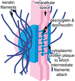
This diagram shows a desmosome. It is made up of a dense
cytoplasmic plaque, to which the intermediate filaments attach.
Desmosomes and Hemidesmosomes
Desmosomes connect two cells together. A desmosome is also
known as a spot desmosome or macula adherens (macula = latin for
spot), because it is circular or spot like in outline, and not belt-
or band shaped like adherens junctions.
Desmosomes are particularly common in epithelia that need to withstand
abrasion (see skin). Desmosomes
are also found in cardiac cells, but the intermediate filament in
this case is desmin, not keratin (which is found in epithelial cells).
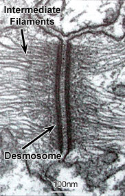
The picture shows an EM of a desmosome formed between two cells.Notice
the phase dense material between the two cell membranes, which is
mad up of transmembrane linker glycoproteins (e.g. demosgleins and
desmocollins - which are cadherin proteins). Also notice the intermediate filaments running from the desmosome into the cytoplasm.
Other proteins run across the membrane into the intracellular space,
to connect the two cells together. These 'transmembrane linker'
proteins are called desmoglein and desmocollin, which are types of cadherin.
Hemidesmosomes
These look similar to desmosomes, but are different functionally,
and in their content. The connect the basal surface of epithelial
cells via intermediate filaments to the underlying basal lamina.
The transmembrane proteins of hemidesmosomes are not cadherins,
but another type of protein called integrin.
This electron micrograph shows a Hemidesmosome (H), and two of
the three layers of the underlying basal lamina. LL - lamina densa,
LD - lamina lucida. Integrins in the plasma membrane link the cell
to the extra-cellular matrix.
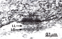
from Wheater's Functional Histology.
Gap Junctions.
Gap junctions are the most widespread of all cell junctions in
animal tissues. Gap junctions thus couple cells electrically and
metabolocially, enabling cells to communicate with each other directly.
Gap junctions can open and close in response to changes in calcium
levels, and pH. Gap junctions form in a narrow gap of 2-4nm, between
two adjacent cells.
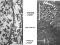
The picture shows an EM (A) of two gap junctions between two cells,
and a freeze fracture EM (B) of the particles on the cytoplasmic
face of the plasma membrane.
A
group of protein molecules called connexins form a structure called
a connexon (each particle in B above is a connexon). When connexons
(blue in the diagram to the right) from two adjacent cells (red
and yellow in the diagram) align, they form a continuous channel
between them.
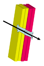
This channel is big enough to allow small molecules such as inorganic
ions, and other small water soluble molecules (smaller than 1000kDa)
to pass between the cells. However the channel is too small for
proteins, nucleic acids or sugars to pass through.