Meiosis
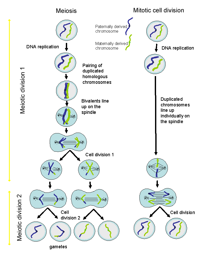
The diagram on the left shows meiotic cell division. That on the right shows mitotic (normal) cell division. Meiosis is from the greek work for 'diminuition'.
- A diploid nucleus contains two pairs of each type of chromosome (autosomes) together with the sex chromosomes (X and X, or X and Y).
- One of these chromosomes is derived from the male parent (parental chromosome) and one from the female (maternal chromosome).
- The chromosomes in this pair are called homologs - there is one paternal and one maternal homolog.
Meiosis |
Mitosis |
|
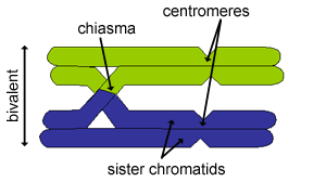
Homologous chromosomes duplicate to form sister chromatids, as in mitosis. The arms of sister chromatids are glued together by a protein called 'cohesin'. However, in meiosis, the two pairs of chromosomes (paternal and maternal) stay together on the spindle of the equator to form a bivalent (contains 4 chromatids) |
Homologous chromosomes duplicate, but remain separate. | |
In the long prophase of meiotic division 1, genetic material can 'cross over' between maternal and paternal pairs of chromosomes (non-sister chromatids). On average, 2-3 cross-overs per chromosome occur for humans. Each cross-over between two non-sister chromatids is called a chiasma (plural 'chiasmata') This exhange of genetic material between maternal and paternal chromosomes is known as 'genetic recombination' Each cross-over between two non-sister chromatids is called a chiasma (plural 'chiasmata')
These random cross-over events scrambles the genetic material of each of the chromosomes in the gametes, helping to produce diverse offspring. The chiasmata are also important in holding the maternal and paternal homologs together until they are separated at anaphase 1. |
Cross-overs do not occur between non-sister chromatids, as they are separate. | |
At anaphase of meiotic division 1, the maternal and paternal chromatids are separated, such that one daughter cell inherits one mostly paternal homolog, and the other one mostly maternal homolog. |
At anaphase one of each pair of chromosomes goes to each daughter cell, such that each daughter cell inherits one copy of the paternal chromosome, and one copy of the maternal chromosome - as in the diagram on the right hand side. | |
The resulting cells after meiotic division 1 are diploid - but unlike mitotic divisions: |
||
Cells then enter another round of cell division, without DNA duplication - meiotic division 2, to form a gamete. |
The resulting cells are diploid. 2 diploid cells are produced. |
In humans, which have 23 pairs of chromosomes, as a result of the random mixing, 223 = 8.4 x 10⁶ genetically different gametes can be produced. Because there is some swapping (or recombination) of genetic material between the pairs of homologous chromosomes, the actual number of genetically different gametes can be very much higher.
Sometimes, homologous chromosomes do not separate normally in meiosis 1, (nondisjunction). As a result, embryos can form that either have extra copies, or lack copies, of one or more chromosomes. Many of these embryos die, but in the condition 'Down's syndrome', there is an extra copy of chromosome 21 (X-chromosome). An extra copy of an autosomal chromosome is known as 'Trisomy'.
In formation of the female gametes (oocytes), only one ovum is produced as a result of meiosis. After the first meiotic division, one of the daughter cells is larger, and is able to form into an ovum (secondary oocyte), and the second cell is small (the first polar body) and cannot produce an ovum. Similarly, after the second meiotic division, one of the daughter cells is large (ovum) and the other is small (second polar body) and cannot produce an ovum.
Follow these links to find out more:
Oocytes
Sperm
Fertilisation
Early development of the mouse embryo