Hepatocyte function
The hepatocytes (epithelial cells of the liver)
form branching plates of cells, often only one cell thick, between
a system of capillary sinusoids that connect the portal tracts to
the central vein.
To facilitate the exchange of a wide variety of substances between
the blood and hepatocytes,the hepatocytes are directly exposed to
the blood passing though the organ, by being in close contact with
the liver blood sinusoids. The sinusoids carry blood from the edges
of the lobule to the central vein.
The sinusoids are lined by two types of cell 1) phagocytic cells
called Kupffer cells (macrophages), which phagocytose
dead red blood cells, particulate matter, and micro-organisms. 2) sinusoid lining cells which are similar to the
endothelial cells that line other blood vessels.
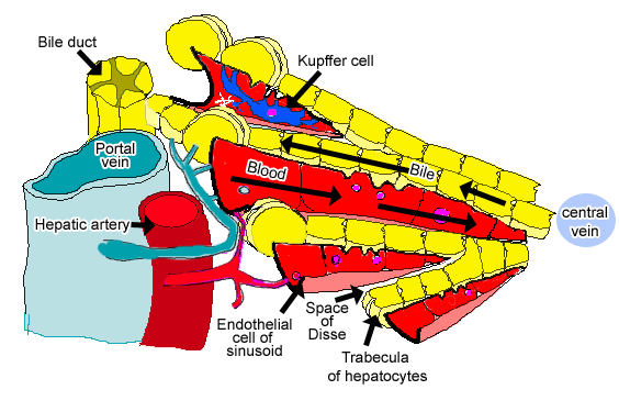
The blood enters through portal tracts at the outer edge of the liver lobule, and filters through the sinuisoids
which are in close connection with the liver hepatocytes, until
it reaches the central hepatic vein, where it drains
away. Thus the flow of blood is from the outside to the inside of the lobule.
In contrast, bile flows through small canaliculi formed by the hepatocytes themselves, and it flows from the inside of the liver lobule towards the outside.
This diagram shows how the relationships between the hepatocytes
and the liver sinusoids.
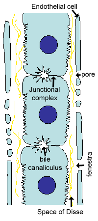
The endothelial cells of the liver sinusoids (capillaries) have
pores, including large pores called fenestra. Furthermore
the basement membrane is discontinuous and non-obstructing.
This allows blood plasma to enter a space between the endothelial
cells and the hepatocytes, called the space of Disse (perisinuisoidal
space). Blood cells and platelets are excluded. The hepatocytes
have many microvilli which project into this space,
to increase absorption from the plasma.
The space of Disse also contains lipocytes,
that store fat, and vitamin A. They can also become contractile,
and they make the type III collagen fibres (reticular fibres) also
found in the space of Disse.
Hepatocytes synthesise and secrete bile into a
system of tiny bile canaliculi which are present
between adjacent hepatocytes. These canaliculi do not have a duct-like
structure but consist merely of localised enlargements of the intercellular
space between adjacent cells. These canaliculi empty into short
canals of Hering close to the portal area, which
then open into duct cells in a bile ductule in the portal area.
This electron micrograph shows two hepatocyte cells which
contain mitochondria and microvilli, together with the space
of Disse, and the endothelial cell of the sinusoid.
See if you can identify them for yourselves.
As well as mitochondria, the hepatocytes contain extensive rER
and Golgi. Proteins are secreted into the sinusoids.
The smooth ER is where damaging compounds such
as steroids, alcohol, drugs and toxins are converted or detoxified.
Stored glycogen is also metabolised the the smooth ER. The hepatocytes
also take up haem, the breakdown product of erythrocytes,
produced by the Kuppfer cells. The hepatocytes
convert this to bilirubin, an iron depleted form
of haem. The sER conjugates bilirubin to glucoronic acid, to make
it water blood and some tissues, giving the skin a yellow colour
known as jaundice.