Female Reproductive System: Early embryonic development
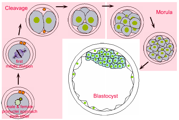
Cleavage involves rapid mitosis, with synthesis of DNA, but no increase in cytoplasmic mass, such that the cells re-obtain the normal ratio of nucleus to cytoplasm.
The first cleavage results in two equally sized cells.
At the 8 cell stage, the cells change their shape, become polyhedral, and the cells fit together tightly - this is called compaction. The cells now start to form gap junction and tight junctions between them.
Cleavage now forms cells that are less regular in size, to form a morula of 16-32 cells. There are now two layers of cells - and inner layer and an outer layer. Only the outer layer of cells contain tight junctions between them.
Fluid filled spaces start to appear between the cells in the inner layer, and coalesce to form the blastocytic cavity.
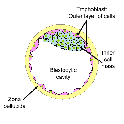
This diagram shows the blastocyst in more detail.
The outer layer of cells is the trophoblast. These cells are connected by tight, gap and adhering junctions.
Inside the trophoblast, there is an eccentrically placed inner cell mass called the embryoblast, and the blastocytic cavity. The region where the embryoblast contacts the trophoblast is called the embryonic pole.
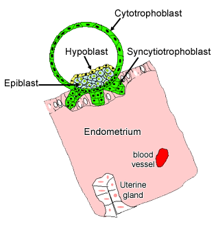
The blastocyst then 'hatches' out of the Zona pellucida and attaches to the endometrium.
Just before implantation, the cells in the embryoblast start to differentiate into two layers - the epiblast (primary ectoderm), and an internal layer of cuboidal cells called the hypoblast (or primary endoderm). A basal lamina is laid down between these two layers.
The resulting two layered embryoblast is called the bilaminar germ disc.
After gastrulation, the epiblast becomes the ectoderm (secondary ectoderm)
At the end of the first week:
The blastocyst adheres to the uterine wall. When the trophoblast comes into contact with the endometrium, the trophoblast at the embryonic pole proliferates. Some of the proliferating cells fuse to form a mass of cytoplasm with multiple nuclei - and this is known as the synctiotrophoblast.
The cells forming the wall of the blastocyst retain their cell membranes and are called the cytotrophoblast.
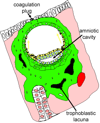
By 9 days, the embryo is completely implanted into the uterine endometrium.
Fluid has collected between the cells of the epiblast (blue), and coalesced to form the amniotic cavity. The cells that form the amniotic membrane are called amnioblasts, and the amniotic membrane separates the new cavity from the cytotrophoblast.
Meanwhile, cells migrate out from the hypoblast (yellow) to line the blastocyst cavity, forming a primary yolk sac, and then later they form a secondary, or definitive yolk sac.
Until day 9, the embryo can rely of diffusion for nutrients and removal of waste. The placenta begins to form on day 9, as trophoblastic lacunae open within the synctiotrophoblast. Maternal capillaries near by, expand to form maternal sinusoids, that anastomose with the trophoblastic lacunae. The cytotrophoblast grows out into these blood filled lacunae - forming primary stem villi and eventually forming blood vessels that connect with blood vessels in the embryo by the end of the third week.
The details of cell migration between days 9 and 13 are not important for you to know.
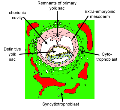
By day 12, the space that was the blastocyst cavity, has now become the secondary, or definitive yolk sac.
The extra-embryonic mesoderm (derived from the epiblast) forms the outer layer of the yolk sac wall, and is important in blood formation (haematopoesis).
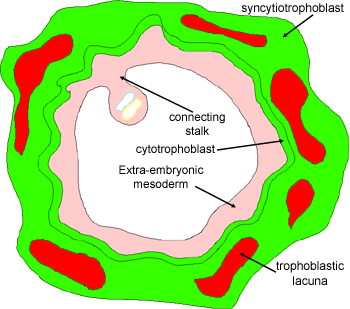
By the end of the second week, the bilaminar germ disc is suspended in the chorionic cavity by a thick connecting stalk.
The major event of the third week is gastrulation.