In this low power image of the pituitary gland, can you identify
the paler staining posterior pituitary, and the darker staining
anterior pituitary. The posterior pituitary is connected to the
hypothalamus by the pituitary stalk.
The pituitary (also known as the hypophysis) is found
at the base of the brain, about 1cm in diameter, lying beneath the
third ventricle in a bony cavity (sella turcica) in the base of
the skull. It has a complex structure. This diagram of the pituitary
shows its main features.
The anterior and posterior parts of the pituitary have different embryological origins.
This picture of the pituitary has been stained with PAS and orange
G. You should be able to identify the anterior and posterior pituitary.
(The brightly stained part is the anterior). Rathke's pouch (vestiges
of) is stained bright purple, by this stain.
The posterior part of the pituitary has its
embryological origins in nervous tissue. It is
formed from a downgrowth of the diencephalon that forms the
floor of the third ventricle.
The anterior part is derived from an upgrowth from the oral ectoderm of the primitive
oral cavity called Rathke's pouch.
Along the posterior part of the the anterior lobe there is a narrow
region called the pars intermedia, which also has
its embryological origin in Rathke's pounch. The pars intermedia
is poorly developed in humans. The glandular epithelial part of
the pituitary is also called the adenohypophysis - (anterior pituitary,
pars intermedia and pars tuberalis).
These different origins are reflected in the histological structure
of the anterior and posterior parts of the pituitary.
This is a magnified image of the anterior pituitary, stained with H&E showing two
types of chromophils; pink - acidophils, which
are more common, and purple - basophils. (Occasionally,
you may be able to identify a third type, which are poorly stained,
these are resting/degranulated chromophils)
Anterior Pituitary
You should also be able to see two types of chromophils (cells
which take up stain) called acidophils and basophils.
Acidophils and basophils are
divided into different classes of cells which have different secretory
products and target organs. The subtypes of acidophils and basophils,
are described in the table below. These cannot be distinguished
by H&E or other histological stains, but can be stained specifically
by immunohistochemical techniques.
The anterior pituitary secretes five different types of hormone.
See the table below.
The capillaries in this gland are fenestrated, to enable passage
of hormones from the secretory cells into the bloodstream.
Glandular Disorders and diseases
The anterior pituitary also contains one type of chromophobe
(cells which stain only weakly) These are resting/degranulated chromophils.
|
Type of chromophil |
Class |
Secretory Product |
Target Organ |
|
Acidophil
|
Somatotrophs (about 50%) |
growth hormone (GH, also known as somatotrophin,
STH) |
General, but a major target is the chondrocytes in epiphyseal
growth plates. Promotes growth (together with insulin like growth
factors) |
| Mammotrophs (about 20%) |
prolactin (PRL) |
Milk producting tissue (alveolar cells) in the breast |
| Basophil |
Corticotrophs (about 20%) |
ACTH (Adrenocorticotrophic hormone,) also known as corticotrophin.
Melanocyte-stimulating hormone (MSH) |
Trophic (trophic - from the greek - to nourish)-
general action - promote growth and secretory activity in other
endocrine glands.
ACTH target is the corticosteroid cells of the adrenal cortex.
MSH target is the melanocytes.
TSH target is the follicular epithelial cells of the thyroid.
FSH target is the follicular cells of the ovaries, to promote growth
of follicles, or the sertoli cells of the testes to promote spermatogenesis.
LH target is developing follicles - promotes ovulation, or the
Leydig cells of the testes - where it promotes secretion of adrogenens,
aiding spermatogenesis.
Three of these trophic hormones (TSH, FSH and LH) are glycoproteins,
so they can be stained up by the technique PAS. |
| Thyrotrophs (about 5%) |
TSH (thyroid stimulating hormone), also known as thyrotrophin |
| Gonadotrophs (about 5%) |
gonadotrophins: FSH (follicle stimulating hormone) and LH (Lutenising
hormone). |
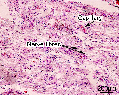
Posterior Pituitary.
The posterior pituitary looks very different to the anterior pituitary (see adjacent image).
It contains non-myelinated axons which are the neurosecretory cells.
The cell bodies of these cells are located in the hypothalamus.
It only secretes two hormones:
Antidiuretic hormone (ADH) which acts on the
kidney, and oxytocin, which acts on the uterus.
Find out more about hormones.