This eMicroscope is a section through part of the spleen. Can you identify the germinal centres, white and red pulp, arterioles, other blood vessels and the capsule.
This image may also be viewed with the Zoomify viewer.
The spleen is the largest mass of lymphatic tissue in the body, and is found between stomach and diaphragm. Like the lymph nodes, it also has a hilus (hilium) which is where the major blood vessels enter and leave. Like the thymus, it only has efferent lymph vessels, which leave from the hilium, and it does not have afferent lymph.
As well as acting as a store for platelets, it has two main functions:
This eMicroscope is a section through part of the spleen. Can you identify the germinal centres, white and red pulp, arterioles, other blood vessels and the capsule.
This image may also be viewed with the Zoomify viewer.
There two main types of tissue in the spleen are specialised for its two main functions:
White pulp contains lymphoid aggregations, mostly lymphocytes, and macrophages which are arranged around the arteries. The lymphocytes are both T (mainly T-helper) and B-cells.
Red pulp is vascular, and has parencyhma and lots of vascular sinuses. These are sinuosoids - a specialised type of capillary, which is very leaky.
The lining endothelial cells have wide slits between their lateral margins, that act as a filter. The blood cells have to move through these slits, before they can leave the spleen and worn out, or defective blood cells are damaged during this process. The damaged cells are then phagocytosed by the numerous macrophages in the red pulp, that lie just next to the sinusoids.
The spleen is covered by a dense capsule, and there are connective tissue trabeculae, which provide internal support for the spleen, and carry the blood vessels into the spleen.

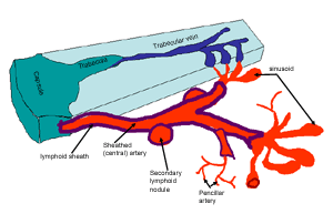
This diagrammatic representation of the spleen, should help you understand where red and white pulp come from.
It shows how the artery has a lymphoid sheath surrounding the artery, as it enters the spleen, with aggregations of secondary lymphoid tissue. This is where the regions of white pulp are found.
Then as the artery loses its sheath and branches to form pencillar arteries, and sinusoids, making up the red pulp.