Male Reproductive system: Epidymis
The genital ducts
The looped seminiferous tubules in the testes
are connected to the genital duct system which
transports the spermatozoa and fluid component of the semen to
the outside.
This duct system is made up of the tubuli recti (short straight tubules connected to the seminiferous
tubules), the rete testis - which is found in
the mediastinum testis. The rete testis
empties into the ductuli efferentes that lead into the ductus epididymus.
The ductus epididymus empties into the vas deferens, which empties into the ejaculatory
duct, which empties into the urethra and passes to the outside.
The arrangement of these ducts is shown in the diagram.
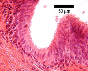
This is an image of the epididymis.
Can you identify the specialisations on the lining epithelium?
Is this a simple, stratified or pseudostratified epithelium? Can
you also identify the basement membrane, and layer of smooth muscle
surrounding the duct?
The epithelium also contains 'basal' cells - the nuclei are close
to the basement membrane - can you identify these.
The epididymis
The ductulis efferentes and the ductus
epididymus make up the epididymis. These
ducts are highly coiled, and make up a single tube in the epididymis
that can be up to 6 metres long. This means that several sections
of it can be found on a single slide.
The ductus epidymis is a storage reservoir for
spermatozoa that become matured here, and competent to fertilise
ova. Fluid is absorbed here, and the epithlium is also secretory.
The epithelium is pseudostratified, with long immotile 'sterocilia'
which are in fact very long microvilli. These are thought to be
involved in absorption of fluid. The cells are also thought to
be secretory, but the nature of the secretions is unknown.
Vas (ductus) deferens
This image shows the vas deferens
(ductus deferens). It is a thick walled tube,
lined with an inner (I) and outer layer of longitudinal smooth
muscle (0) and a middle layer of circular muscle (C). Can you
recognise these three layers of muscle?
Stimulation by the sympathetic nervous system causes
the contraction of this muscle, to expel its contents into the
urethra during ejaculation. The duct is lined by a pseudostratified
epithelium, and the supporting lamina propria is folded, which
means it is able to expand during ejaculation.
The distal portion of ductus deferens (the ampula)
receives a duct draining from the seminal vesicle
- forming the short ejaculatory duct. These ducts converge to
join the urethra, as they pass through the prostate gland.
This image shows the epithelial lining of the vas deferens.
Can you identify the pseudostratified tall columnar cells?
It is similar to that of the seminal vesicles.