The human mouth is for
- Breaking up, eating, moistening, and tasting food,
- Speaking,
- Facial expression,
- Breathing
The human mouth is for
This diagram shows across section through the lip and tooth, showing some of the main features of these structures.
All of the oral mucosa is made up of a thick stratified squamous epithelium, supported by a lamina propria. The epithelium is thick because the epithelial lining of the oral cavity is subject to a lot of wear and tear.
In mobile areas, such as the soft palate, underside of the tongue, floor of the mouth, and mucosal surfaces of the cheeks and lips, the epithelium is not keratinised.
In other areas, such as the gums (gingivae), hard palate, and most of the upper surface of the tongue, the epithelium is keratinised.
Underneath the oral mucosa, there is a tough collagenous submucosal layer, with accessory salivary glands, except where the oral mucosa lies over bone, where the submucosa is thin.
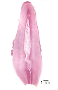
This is a photograph of a section through the whole lip.
Look at this picture of the lip, and identify the vermilion border, accessory salivary glands, hairs, oral mucosa (mucosa lining the mouth), and muscle. (Scale bar is 1 mm). The accessory (or minor) salivary glands are seromucous.
 How is the epithelium of the oral mucosa different to that of the vermilion border?
The inner surface has thick stratified squamous epithelium.
The outer surface has thin, lightly keratinised epithelium
Why do you think the vermilion border is normally red (in the living). Hint - this is more to do with blood supply than with lipstick!
The vermilion border is red, because it is highly vascularised.
The tooth can be divided into two main areas: the crown and the root.
Most of the hard tissue in teeth is dentine, a special calcified tissue, derived from mesenchyme. The dentine in the root is covered by a layer of cementum, calcified tissue derived from mesenchyme. The tooth is then connected to bone by the periodontal ligament, which has wide bundles of collagen fibres, and is embedded in bony ridge called alveolar ridge.
Teeth is made up of three layers, enamel, dentine and a pulp cavity. The crown is protected by layer of enamel, a very hard, highly mineralised tissue, which is derived from ectoderm. Cementum, dentine and enamel differ from bone, in that they are not vascularised. Enamel also does not have collagen as its main consituent. It is made up of crystals or prisms of calcium phosphate. The centre of tooth is made up of a pulp cavity that extends down through the roots as a root canal. This region contains the nerve and blood supply to the tooth.
Gums, or gingiva is the name for the oral mucosa that covers the tooth. At the gingival crevice (or sulcus), the cells in the epithelium of the gum adhere to the tooth enamel via a basement membrane. If bacteria calcify here, and accumulate, they can disrupt this seal, and the periodontal tissues can become infected and inflamed.
Dentine is made by odontoblasts, that lie on its inner border. Production of dentine is limited to the pulpal surface. About 90% of dentine is type I collagen, and about 70% of wet weight is hydroxyapatite.
Odontoblasts are tall columnar secretory cells. Their secretory processes are embedded in the matrix, which is impregnated with parallel dentine tubules. Dentine is laid down, and then calcified. Thus there is a thin layer of 'pre-dentine' which is not calcified between the dentine and odontoblasts.
Enamel is made by ameloblasts, tall columnar ectodermally derived cells. It is produced before the tooth erupts. Each ameloblast has an elongated tip called a Tomes process, that secretes the organic matrix of an enamel rod. 96% of a tooth is mineralised. As the ameloblasts die when the tooth errupts, the enamel layer cannot be repaired.
In this low power image of an adult tooth, can you recognise the enamel, dentine, odontoblasts and pulp cavity in this section of an adult tooth?
Look at this magnified section through a tooth, and identify the layers of dentine, predentine, odontoblasts, a relatively cell -free layer called the cell free Zone of Weil and the dental pulp.