Oral tissues: Tongue
Anterior surface of the tongue.
The tongue is a mass of interlacing skeletal muscle , connective tissue with some mucous and serous glands, and pockets of adipose tissue, covered in oral mucosa.
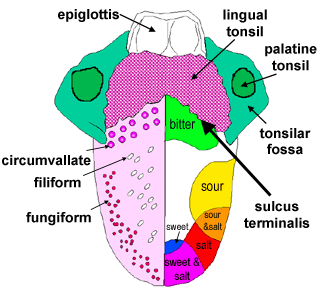
A V-shaped line (shallow groove)- the sulcus terminalis, divides the tongue into an anterior 2/3 and a posterior 1/3.
The mucosa covering the upper surface of the tongue is thrown into numerous projections called the lingual papillae in the anterior 2/3rd of the tongue.
In the posterior 1/3rd, there are no papillae, but there are lots of lymphoid follicles present.
The three types of papillae are:
- fungiform (mushroom like)
- filiform (filum - thread like)
- circumvallate
(foliate papillae are uncommon in humans)
Toggle labels
This image may also be viewed with the Zoomify viewer.

This photograph shows a vertical section of tongue, that contains both filiform and fungiform papillae. See if you can identify them, and notice how the epithelium for the two different types of papillae are different.
Fungiform and Filiform Papillae
The slide above shows a vertical section through the tongue, taken close to its tip.
Can you identify the fungiform and filiform papillae, glands, and skeletal muscle.
Notice the type of epithelium that covers the superior surface of the tongue (including the papillae).
Use the information below, and the diagrams to help you.
Filiform papillae are the most common. They are keratinised and in the living, they look white.
Fungiform papillae are not keratinised, but are highly vascularised. In the living, they look red. most of the fungiform papillae have some taste buds.
Underneath the papillae, there are mucous and serous glands, pockets of adipose tissue, and a layer of skeletal muscle and connective tissue. The skeletal muscle is arranged in three different planes, which allows the tongue to perform a number of complex movements.
Circumvallate papillae
Now have a look at this section, a vertical section of tongue taken
just anterior to the sulcus, that shows part of a circumvallate
papilla . See if you can identify Von Ebner's glands, the cleft
of the circumvallate papilla, the taste buds, and the muscle layer.
The papillae in this region are very large, notice the scale bar.
These papillae have taste buds in the medial walls of the cleft.
These papillae are larger than the other two types of papillae.
Glands, called Von-Ebner's glands (serous glands) open into
the cleft.
Taste buds contain epithelially derived taste receptor cells, sustentacular
cells that surround a small central cavity tat opens onto the surface
as a tiny taste pore. Basal cells renew both of these types of cell.
Chemicals stimulate the receptors, and initiate impulses in the
afferent nerve fibres.
A high resolution version of this image (without labels) may also
be viewed with the Zoomify viewer.