Nails
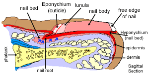
The distal end of each digit is protected by a strong plate of hard keratin, called a nail or nail plate, which grows out from a nail bed. The nail bed, is a specialised form of skin epithelium, and has the same four layers of the epidermis of skin, with the nail plate being analogous to the stratum corneum layer.
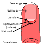
The nail plate is made up of tightly packed, hard, keratinized epidermal cells. It has a nail body, a free edge, and extends deep into the dermis at the proximal end to form the nail root (or nail groove).
The proliferating cells in the nail root form the nail matrix, and their proliferation (in the stratum basale) make the nail elongate continuously (blue arrows). As the cells here approach the dorsal surface of the nail, they are displaced distally (in the direction of the blue arrows), and are gradually transformed into hard keratin, causing the nail plate to lengthen and strengthen. This layer is thin enough for colour to show through from the vascular dermis below.
The white crescent at the proximal end is called the lunula. It is white because the underlying epithelium is thicker here, and the colour of the dermis does not show through from underneath.
The epithelium underlying the nail bed and nail plate, forms a continuous fold, first forming the cuticle or eponychium (epo=above) at the proximal end of the nail, overlying the nail plate , then the nail bed underneath the nail plate and finally the hyponychium (hypo=below); a thickened region of stratum corneum that secures the nail to the finger tip, and lies below the nail plate.
This photograph shows an H&E section through a finger (sagittal), but the nail itself has been removed.
Can you identify the epidermis and dermis, phalanx, eponychium, hyponychium, and nail root?
The photograph on the right hand side shows a transverse section through a nail. Can you identify the prominent epidermal ridges?
In this section, the nail is present, can you identify it?.