Epithelia: Specialisations
Cilia
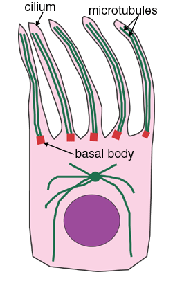
Cilia are hair-like motile processes. They are about 10µm long and about 0.2µm in diameter, and are longer and thicker than microvilli. They are made of microtubules - shown here in blue. There is a ring of doublet microtubules, and a central pair of singlet microtubules. (See cross-sectional diagram below). At the base of the cilium is the basal body, into which the microtubules are anchored.
You can see that arms made of dynein link the doublet microtubules. Dynein is a microtubule motor protein. When the dynein molecules attach to their adjacent doublet microtubule, this makes the cilium bend, via a sliding motion between the microtubules. When dynein releases, the cilium straightens up again. This cyclical action causes the cilia to beat. Dynein uses ATP for energy.
This is a cross sectional view of a cilium.
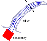
This diagram shows an LS through a cilium, showing the location of the basal body and microtubules. The dotted line is where the cross section is taken for the diagram opposite
Can you can identify the ciliated cells of the trachea.
The beating of the cilia on cells is used to propel mucus along the trachea, for example.
Microvilli
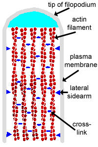
These are small finger like projections, about 1
µm in length, and 90nm or so in diameter. The microvilli are shorter and narrower than cilia. (Microvilli is the plural of microvillus.)
They contain bundles of parallel actin filaments held together into a bundle by cross-linking proteins called villin and fimbrin.
Lateral arms containing myosin I and calmodulin link the actin filament bundle to the plasma membrane.
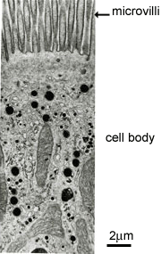
Microvilli are present on the luminal surface of many epithelia, particularly those specialised for absorption.
Microvilli increase the area of the free surface of the epithelium available for absorption.
This picture shows a Transmission EM through a tall coumnar cell in the small intestine. At the top of the cell, that projects into the gut lumen, there are the fine structures called microvilli.
Because this is a section through the cell, it does not cut perfectly symmetrically through all the microvilli in the section, and this explains the appearance of the microvilli as shown here. (Think about it!).
This is an H&E section of thick skin. The epithelium has many layers of cells, and has a thick layer of keratin (stained red) on the top (apical) surface.
Can you identify the basal layer, keratin, and epithelial layers?
The epidermis is folded, and dermal papillae are found where the dermis projects up into the folds. Can you identify a papilla?
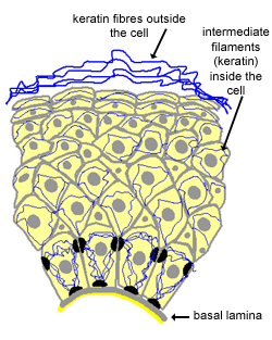
Keratin is a type of intermediate filament found in cells.
In the skin, basal cells (next to the basal lamina) divide and move out into the layers above. (For more detail, see the section
on skin). As they reach the outer layers, they start to lose their nuclei and cytoplasmic organelles, the cells degenerate and become 'keratinized squames' of the outer keratinized layer.
They finally flake off from the surface (and are a major constituent of household dust).


