Muscle: Skeletal and Cardiac Muscle Ultrastructure
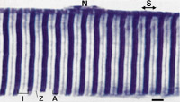
This is a high power, light micrograph of a muscle fibre showing
the banding pattern. There are light stripes - which are called
the 'Z' lines, and darker wider stripes called the 'A' bands. (A
- for anisotropic - because in a polarizing light microscope, the
dark bands are birefringent)
The Z-lines are midway inside a light band, called the 'I' band.
(I for isotropic - because in a polarising microscope, these bands
are much less birefringent than the A bands).
The muscle sarcomere is the repeating unit (S) between two Z-lines.
N - is a nucleus on the outside of the fibre.
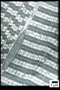
This is an electron micrograph (EM) of a skeletal muscle fibre.
(The sarcomeres of cardiac muscle have a very similar organisation).
Notice how the stripes appear less regular than in the light microscope.
This is because the repeating muscle sarcomeres are arranged in
longitudinal structures called 'myofibrils' (these run from top
right to bottom left of the picture).
This is the same EM with labels to show the organisation of the
muscle sarcomeres. Z-lines mark the boundaries of the sarcomeres.
The dark staining region in the centre of the sarcomere is called
the A (anisotropic) band. The lighter staining band, through which
the Z-line passes is called the I (isotropic) band.
A diagram of a muscle sarcomere is shown below.
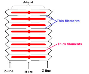
Thin filaments, which consist mostly of the proteins actin, troponin and tropomyosin, insert directly into the Z-lines.
Thick filaments consist mostly of the proteins myosin and titin. Titin connects the thick filaments to the Z-line.
There are about 300 molecules of myosin in each thick filament,
and at the end of each molecule are globular heads that stick out
and bind to actin in the thin filament. When the muscle is stimulated,
the heads bind to actin, and pull on actin to pull the thin filaments
towards the M-line, increasing the overlap between thick and thin
filaments, and making the muscle shorten. This uses ATP for energy.
In relaxed muscle, troponin/tropomyosin stops myosin from binding to actin. When muscle is stimulated,
calcium is released, binds to troponin, and this then allows myosin to bind to actin.