Mitosis
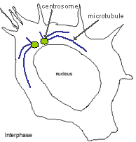
Interphase: (between cell division). Here the cell is about to start entering mitosis. The centrosome (green) has duplicated. Microtubules (blue) grow out of the centrosomes. The centrosome and microtubules cannot be seen in the phase picture below, but are shown in the diagram, to show how these structures re-organise during mitosis.
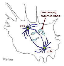
Prophase - the start of mitosis
Nuclear envelope breaks down, and chromosomes condense (light blue structures in the diagram opposite). Microtubules re-organise to form mitotic spindle. The centrosomes move to opposite ends of the nucleus to form the spindle poles.
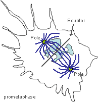
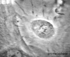
Prometaphase
Duplicated chromosomes (light blue in diagram opposite) move to the centre of the mitotic spindle (the 'equator'). See if you can identify the chromosomes in the phase micrograph below - they look black and are close to the equator.
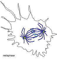
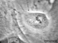
Metaphase
Duplicated chromosomes (light blue) are aligned on the centre (equator) of the mitotic spindle. Notice how the whole structure has rotated, so that the poles are rotated by about 90 degrees compared to that in prometaphase.
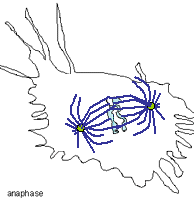
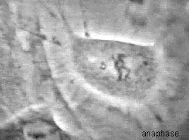
Anaphase
Chromosomes move to the poles (shown in green) of the spindle.
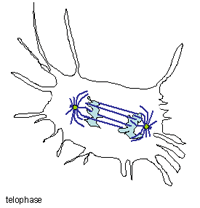
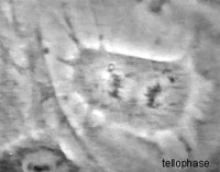
Telophase
Chromosomes have moved towards the poles, they will now start to decondense, and intact nuclei will start to reform.
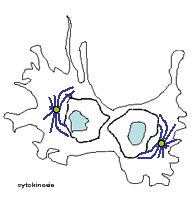
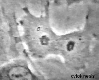
Cytokinesis
Cell is pinched in two, to form two new cells.
The whole of mitosis lasts about 30 - 60 minutes in real time, here it is shown speeded up.
Have a look at dividing cells in a photo.