What is the nucleus?
The nucleus is found in the middle of the cells,
and it contains DNA arranged in chromosomes. It is surrounded by
the nuclear envelope, a double nuclear membrane (outer and inner),
which separates the nucleus from the cytoplasm. The outer membrane
is continuous with the rough endoplasmic reticulum. The nuclear
envelope contains pores which control the movement of substances
in and out of the nucleus. RNA is selectively transported into
the cytoplasm, and proteins are selectively transported into the
nucleus. The nuclear membrane is supported by a meshwork of intermediate
filaments, called nuclear lamins.
One or more darkly staining spherical bodies called the nucleoli are
found inside the nucleus. These are the sites at which ribosomes
are assembled. Nucleoli are most prominent in cells that are synthesising
large amounts of protein.
Most cells have a single nucleus, though some have none (ie. red
blood cells), and some have several (i.e. skeletal
muscle).
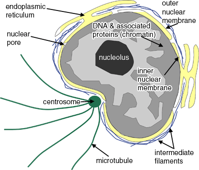
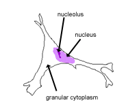
This image below shows a diagram of the nucleus.
Nuclei look different cell types, and when cells divide. For example, in different types of white blood cells, in interphase, the nucleus can have one, or more lobes, and the number of lobes is characteristic of the type of white blood cell.
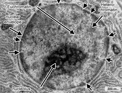
This picture shows an electron micrograph of a nucleus. The short
white arrows are pointing to nuclear pores. Note the appearance
of eu- and heterochromatin, and the nucleolus. Heterochromatin stains
more densely than euchromatin, but they are both forms of chromatin.
Chromatin is the name for the diffuse granular mass of DNA found
in interphase cells.
Heterochromatin is less abundant, relative
to euchromatin, in the large nuclei of active cells
than in the small nuclei of resting cells, such as small lymphocytes.
Euchromatin is "active" chromatin, containing
DNA sequences that are being transcribed into RNA.
The nucleolus is the site in the nucleus where
ribosomal RNA is transcribed. It is then linked to the subunits
of the ribosome, and transported out of the nucleus through nuclear
pores. The ribosomes assemble, and translation of RNA and protein
synthesis occurs in the cytoplasm. Protein synthesis occurs on free
ribosome or on ribosomes attached to the endoplasmic reticulum (rough
endoplasmic reticulum), in which case a pore is formed so that newly
synthesized proteins move into the cisterna of the rough endoplasmic
reticulum. The proteins synthesised on ribosomes attached to the
ER, are then transported to the Golgi, and packaged for secretion.
Types of Nucleus
Cells are normally diploid - this means that
they have a pair - two sets of homologous chromosomes, and hence
two copies of each gene or genetic locus.
However, cells can be haploid, polyploid or aneuploid.
Haploid: only has one set of chromosomes - i.e. in a sperm or
oocyte. Polyploid: Contains more than two sets of homologous
chromosomes. Aneuploid: Have atypical chromosome numbers
- can either have one or two extra chromosomes, or can lose chromosomes.
This is abnormal, and can be diagnostic of cancer.
The karyotype - is a count of how many pairs of chromosomes
there are.
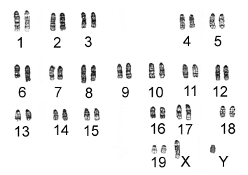 This picture shows a 'G-banded' karyotype from a diploid mouse cell in metaphase. The chromosomes have been arranged into their pairs with their numbers are shown below. The characteristic banding pattern you can see is obtained by staining with Giemsa.
Mice only have 19 pairs plus XY chromosomes, whereas Humans have 22 plus XY.
Have a look at the genome mapping project for humans and other species at the National Center for Biotechnology Information website


 This picture shows a 'G-banded' karyotype from a diploid mouse cell in metaphase. The chromosomes have been arranged into their pairs with their numbers are shown below. The characteristic banding pattern you can see is obtained by staining with Giemsa.