Formation of Ova
Follicular development
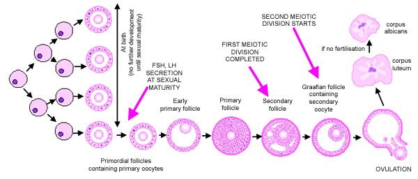
Primordial germ cells multiply during fetal development. At birth,
the ovary contains around 400 000 primordial follicles which contain
primary oocytes. These primary oocytes do not undergo further
mitotic division, and they remain arrested in the prophase stage
of meiotic division I, until sexual maturity (see Meiosis,
in the topic cell). Compare this to male
gametogenesis.
At sexual maturity, two hormones, produced by the pituitary
gland: follicle stimulating hormone (FSH) and lutenising hormone (LH)
cause
these primordial follicles to develop. In each ovarian cycle, about
20 primordial follicles are activated to begin maturation. however,
normally only one follicle fully matures, and the rest contribute to
the endocrine function of the ovary.
When activated, the first meiotic
division is completed. When this happens, the primary follicle has
matured into a secondary
follicle. The second division then starts, and a Graafian
follicle is formed. This contains a secondary oocyte. This second
division is not completed, unless the ovum is fertilised.
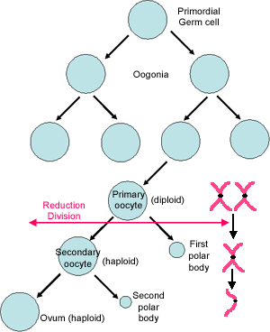
Meiosis
This diagram shows how primary oocytes in the primordial follicles
are diploid, and are starting their first
meiotic division. The pairs of homologous chromosomes
(one maternal, one paternal) pair up on the spindle, and genetic
material can be swapped over. Even the X and Y chromosome (if
present) will pair up, and form a partial bivalent.
At completion of first meiotic division: one
of the chromosome pairs is segregated to each of the daughter
cells. For example, if there is an XY pairing, then one cell will
receive the X, and one the Y chromosome pair.Thus when this division
is completed, the resulting secondary oocyte has 'diploid' DNA, but the chromosome copies are only derived
from one of the original chromosomes in the parent cell. (i.e.
either maternal or paternal). These cells are called 'haploid'.
One of the daughter cells degenerates and forms a 'polar
body'. These are small and degenerate rapidly.
At completion of the second meiotic division:
this division occurs without the DNA being replicated prior
to division. Each daughter cell receives one chromatid from the original chromosome pair to form the gamete producing
cells that only have a single copy of each chromosome. Again one
of the daughter cells forms a polar body.
Thus only one gamete is formed from one primary oocyte (compare
this to male gametogenesis).
For more information compare normal
mitotic division with meiotic division.
Hormonal changes during the menstrual cycle
Development of the follicles is stimulated by production of follicle
stimulating hormone (FSH) by the pituitary gland. Ripening
of the follicles then results in an increase in oestrogen levels,
as oestrogen is secreted by follicular cells. This increase in oestrogen levels
feeds back to the pituitary, and suppresses further release of FSH (negative
feedback). The follicles also release a second hormone called inhibin,
which also suppresses further production of FHS.
As the oestrogen levels rise, this triggers a a mid cycle surge in
a second pituitary hormone called Lutenising hormone (LH),
which causes the follicle to rupture (ovulation).
LH also causes ruptured follicles to lutenise, forming a transitory
endocrine organ called the corpus luteum. This looks
yellow, due to its pigmented lutein cells (luteus is latin for yellow).
The corpus lutein secretes progesterone and oestrogen. The progesterone
levels feed back to the pituitary and suppress further release of
LH. If fertilisation does not occur, the corpus luteum degenerates
into a small white fibrous scar called the corpus albicans. The
resulting decline in progesterone (and to some extent
oestrogen) levels precipitate menstruation. The
decline in oestrogen levels, feeds back to the pituitary and there
is a corresponding increase in FSH to being the cycle all over again.