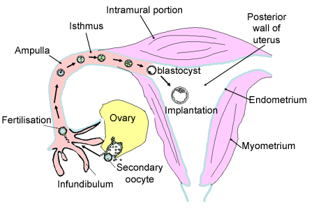
The oocyte is normally fertilised in the fallopian tubes, and by the time it reaches the uterus, 5 days later, it has already formed a blastocyst (see embryogenesis

The oocyte is normally fertilised in the fallopian tubes, and by the time it reaches the uterus, 5 days later, it has already formed a blastocyst (see embryogenesis

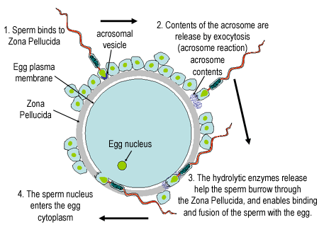
Although many sperm can reach the egg, and bind to it, normally ony one sperm fuses with the plasma membrane of the egg, and injects its nucleus. The surface of the egg is covered with fine microvilli, which interact with the sperm, by clustering, and elongating to hold the sperm firmly in position. A protein called fertilin is important in the subsequent fusion of the egg and sperm plasma membranes, but the exact mechanism is still unclear.
Polyspermy: occurs if more than one sperm fuses with the egg. However, this results in extra mitotic spindles, and faulty segregation of chromosomes, nondiploid cells, and development usually stops.
When the first sperm fuses, there is a rapid depolarisation of the egg, that initially blocks entry of further sperm. This also causes the levels of calcium ions to increase, which initiate the cortical reaction.
The cortical reaction prevents further sperm entry after the egg repolarises, as it results in the proteins in the zona pellucida become 'hardened'.
Once fertilised, the egg is called a zygote.

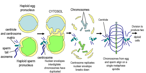
Fertilisation is completed when the two haploid nuclei (pronuclei) come together and combined their chromosomes into a single diploid nucleus (see diagram, adapted from fig 20-34, Molecular Biology of the Cell, 4th edition).
The sperm contributes more DNA than the egg to the zygote, as well as the centriole (Unfertilised human eggs do not have a centriole). The centriole is important for forming the spindle. It replicates to form the first mitotic spindle in the zygote.
After this first division, the zygote then proceeds on to the process of forming an embryo.