Lymphoid: Immune cells
Types of immune cell
This diagram shows the different types of immune cell that are important in the immune system.
- Lymphocytes (B and T).
- Plasma cells - which secrete antibodies, derived from B-cells
- Macrophage - which engulfs micro-organisms and presents antigens on its surfaces to lymphocytes.
- Dendritic cell - presents antigens to lymphocytes on its surface.
- Neutrophil - phagocytic cell
- Reticular cell - also a dendritic cell, which presents antigens.
- Mast Cell - derived from the bone marrow, release histamine, heparin,etc. Have receptors for IgE antibodies on their surface. Involved in allergic reactions.
This diagram shows the two main types of lymphocytes, and the two types of immune responses they are involved in.
B- and T-lymphocytes:
Lymphocytes make up 20-50% of circulating leucocytes.
Most are small (6-9µ),
About 3% are large (about 12-15µ) and have more cytoplasm
- these are mostly activated B-lymphocytes which are on their way
to tissues where they will become antibody secreting plasma cells.
The activated lymphocytes can enlarge to 30 µm in diameter
in lymphoid tissues. They have an intensely basophilic cytoplasm,
due to their increased protein synthesis.
The two types of lymphocytes are:
- B-cells, which are made and matured in the bone marrow
- T-cells, which are made in the bone marrow, but have to migrate and reside in the Thymus before they are matured.
The two types of immune response are:
- Humoral antibody response - B-cells and their
progeny (Plasma cells) make antibodies, helped by Helper T-cells.
Mainly a response to infectious bacteria, and
- Cell Mediated immune response - produced by Killer
T cells, which are cytotoxic. this is primarily a response to infectious
viruses, foreign cells or fungi, which are antigenically different
to the host.
B-lymphocytes.
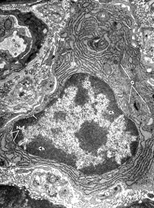
This is an electron micrograph of a plasma cell. You can see that it has lots of Golgi, and rough endoplasmic reticulum, because it is actively secreting immunoglobulin.
Each B-cell is pre-programmed to respond to a particular antigen.
This antigen will stimulate the B-cells, only if they are also stimulated
by cytokines, produced by T-helper cells, that stimulate the B-cells
to divide.
An activated B-cell divides many times and produces many progeny
that have the same antigenicity as the original B-cell. (This is
known as a clone of cells). These B-cells start to secrete antibody.
Some of these B-cell develop into plasma cells,
that are specialised to produce lots of antibody.
The rest remain as small lymphocytes, and persist in secondary
lymphoid organs as long lived memory B-cells, that
can respond quickly when challenged with the same antigen.
T-lymphocytes
T-lymphocytes are formed in the bone marrow,
but mature in the thymus.
Click here to find out more about how
T-lymphocytes are matured in the thymus.
T-cells make up 65 to 85% of the peripheral blood lymphocytes.
They do not make antibodies, and the antigen receptors on their
surfaces are different to those on B-cells. They also have markers
on their surfaces (antigens) called CD antigens (CD for cluster
of differentiation). T-cells start to express these markers during
their maturation in the thymus.The T-lymphocytes also populate the
peripheral lymphoid tissues (lymph nodes, M.A.L.T. and spleen).
There are three types of T-cell:
- Cytotoxic T-cells (killer cells) destroy their
targets by releasing perforin, which inserts into the lipid bilayer
of the target cell, and polymerises into a large membrane channel,
permeabilising the cell, and thereby killing it.
- Helper T cells, which regulate immune responses
by releasing cytokines
- Suppressor T-cells, which downregulate both humoral
and cell-mediated immune responses.
(These last two types are both known as Regulatory T-cells)
Cytotoxic and Suppressor T-cells
both express CD8,
Helper T cells express CD4 antigens.
The different types of T-cell and B-cell cannot be distinuished
using histological stains, but can be distinguished by immunostaining
for the different cell surface markers that these cells express.