Alveoli

This is a cross section through the lung, can you identify alveolar sacs and alveoli?

photo of alveoli

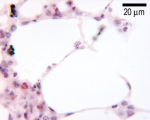
This is a section through the lung at higher magnification. Can you identify the thin type I pneumocytes, macrophages and capillaries?

diagram of alveoli

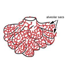
This diagram shows a diagram of an alveolar sac, showing how the organisation of the alveoli, and the network of blood capillaries that surround the alveoli (in red).

The epithelium of the alveoli, contains two main types of cells:

  1. type I pneumocytes: large flattened cells - (95% of the total alveolar area) which present a very thin diffusion barrier for gases.
  2. type II pneumocytes (making up 5% of the total alveolar area, but 60% of cells). These cells secrete 'surfactant' which decreases the surface tension between the thin alveolar walls, and stops alveoli collapsing when you breathe out.

Macrophages are also present.

Can you identify the alveolar spaces, capillary, red blood cell, endothelium, and type I pneumocyte? The dark staining material on the lining of the alveolar space is surfactant.

In this electron micrograph (Mag x 10000), there is a capillary containing a red blood cell (erythrocyte) in the middle of the picture. There are two alveolar spaces on either side. Between the alveoli and the lumen of the capillary are the interalveolar walls or septa that are very thin - about 0.05 µm.

Gaseous exchange takes place through the endothelial cell of the capillary, basement membrane and type two pneumocyte. The wall of the capillary endothelial cell is fused to that of the alveolar cell, and there is only a very thin basement membrane between these two cells. This produces a very narrow gap across which oxygen and carbon dioxide can rapidly diffuse (double headed arrow, in the diagram, with labels on).