Respiratory | Trachea, bronchioles and bronchi
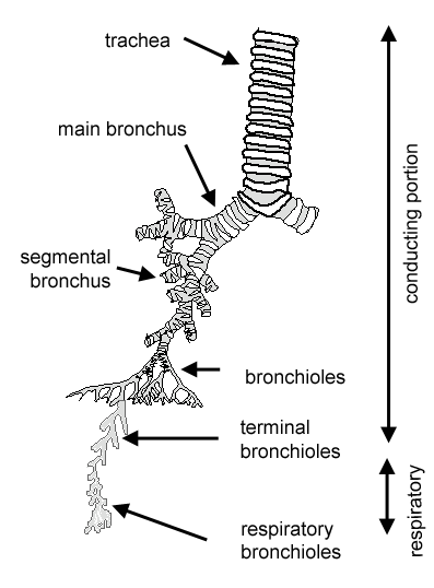
The conducting portion is made up of:
nasal cavities, nasopharynx, larynx, trachea, bronchii and bronchioles
The trachea branches to give rise to two primary
(main) bronchii. These then branch successively to give rise
in turn to secondary and tertiary bronchii.
These then branch to give rise to several orders of progressively
smaller airways called bronchioles, the smallest
of which are called terminal bronchioles. These are the
last components of the conducting portion of the respiratory system.
Terminal bronchioles give rise to respiratory bronchioles, which
ultimately lead to the alveoli.
Find out more about the respiratory portion.
Trachea
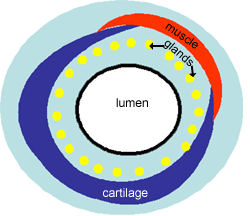
The trachea is a wide flexible tube, the lumen of which is kept
open by 20 tracheal cartilages, which are C-shaped rings of hyaline
cartilage. The gaps between the rings of cartilage are filled by
the trachealis muscle - a bundle of smooth muscle, and fibroelastic
tissue. Together these hold the lumen of the trachea open, but allow
flexibility during inspiration and expiration.
The respiratory mucosa and submucosa are adapted to warm and moisten
the air, and to trap particles in mucous.
Mucosa and sub-mucosa of Trachea
The respiratory mucosa is made up of the epithelium and supporting
lamina propria). The epithelium is tall columnar pseudostratified
with cilia and goblet cells. The supporting lamina propria underneath
the epithelium contains elastin, that plays a role in the elastic
recoil of the trachea during inspiration and expiration, together
with blood vessels that warm the air.
The sub-mucosa contains glands which are mixed sero-mucous glands.
The watery secretions from the serous glands humidify the inspired air. The mucous, together with mucous from the goblet
cells traps particles from the air which are
transported upwards towards the pharynx by the cilia on the epithlium.
This helps to keep the lungs free of particles and bacteria.
This is a cross section through the trachea, showing the major layers.
This is a higher power image of the trachea showing the glands and epithelium in more detail.
Can you classify the epithelium? There are lots of sero-mucous glands in the submucosa layer. The layer of cartilage is not seen here, but instead there is a layer of fibro-elastic connective tissue which runs between the rings of cartilage.
Bronchii
This is a picture of a tertiary bronchus. Compare this picture with that of the trachea
Can you identify the circular layer of smooth muscle, and the cartilage, and some glands in the submucosa?
The smooth muscle is used to control the diameter and length of the bronchii - it contracts during expiration to help expel the air. There is also lots of elastin present in the submucosa, as in the trachea.
The epithelium is now tall columnar, not pseudostratified (difficult to see at this magnification) and has very few goblet cells.
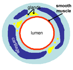
The trachea branches into two primary bronchii, which branch into secondary and then tertiary bronchii. In the tertiary bronchii, there is less cartilage, and it does not completely encircle the lumen, as shown diagramatically above.
Notice also how the mucosa is folded, and think about how this might change as you breathe in and out.
Bronchioles
This is a picture of a bronchiole.
There is no cartilage and no glands. Can you identify the ring of smooth muscle, which is arranged in discrete bundles with a variety of organisations.
The tertiary bronchii branch into bronchioles, which have a diameter
of 1mm or less, and the wall structure changes.
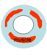
The epithelium is made up of ciliated columnar cells in larger bronchioles, or non-ciliated in smaller bronchioles (difficult to see at this magnification). There are no goblet cells, but there are cells called Clara cells. These cells are secretory - they secrete one of the components of surfactant.
Asthma: because the diameter of the bronchioles is reliant on smooth muscle tone, these airways can almost completely shut if the smooth muscles contract strongly, which can happen in an asthmatic attack.
Terminal Bronchioles.
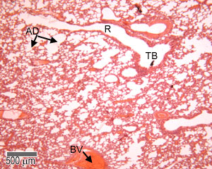
This picture shows a Terminal bronchiole (TB in the diagram). Note that this is at a lower magnification than the three pictures above.This is the last part of the conducting portion of the respiratory system, and has the smallest diameter of all (less than 1mm). There is no cartilage, or glands, some smooth muscle is still present, there are no goblet cells. The epithelium is either columnar or cuboidal.
The final branches of the bronchioles are called terminal bronchioles.
These have a layer smooth muscle surrounding their lumens.
Stimulation of the vagus nerve (parasympathetic) causes the smooth
muscle to contract, and reduce the diameter of the terminal bronchioles.
Small sacs are found extending from the walls of the terminal
bronchii called respiratory bronchioles (R), that are lined by
a ciliated cuboidal epithelium, and some non-ciliated cells called clara cells.
Click here to find out more about the respiratory
portion of the lung.
The respiratory bronchii have a few single alveoli off their
walls.