Skin functions and Layers
Some facts about skin
- Skin is the largest organ of the body.
- It has an area of 2 square metres (22 square feet) in adults, and weighs about 5 kilograms.
- The thickness of skin varies from 0.5mm thick on the eyelids to 4.0mm thick on the heels of your feet.
- Skin is the major barrier between the inside and outside of your body!
Functions of skin
- Protection: it protects against UV light, mechanical, thermal and chemical stresses, dehydration and invasion by micro-organisms.
- Sensation: skin has receptors that sense touch, pressure, pain and temperature.
- Thermoregulation: various features of the skin are involved in regulating temperature of the body. For example sweat glands, hair, and adipose tissue.
- Metabolic functions: subcutaneous adipose tissue is involved in production of vitamin D, and triglycerides.
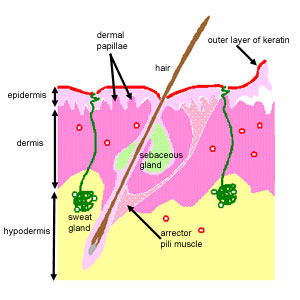
This diagram shows the layers found in skin. There are three main layers: the epidermis, dermis and hypodermis. There are also sweat glands, and hairs, which have sebaceous glands, and a smooth muscle called the arrector pili muscle, associated with them.
Hairs are only found in thin skin, and not in the thick skin present on the fingertips, palms and soles of your feet.
Three layers of skin:
The epidermis: a thin outer portion, that is the keratinised stratified squamous epithelium of skin. The epidermis is important for the protective function of skin. The basal layers of this epithelium are folded to form dermal papillae. Thin skin contains four types of cellular layers, and thick skin contains five. Click here to find out more about the epidermis and its layers.
The dermis: a thicker inner portion. This is the connective tissue layer of skin. It is important for sensation, protection and thermoregulation. It contains nerves, the blood supply, fibroblasts, etc, as well as sweat glands, which open out onto the surface of the skin, and in some regions, hair. The apical layers of the dermis are folded, to form dermal papillae, which are particularly prominent in thick skin.
The hypodermis. This layer is underneath the dermis, and merges with it. It mainly contains adipose tissue and sweat glands. The adipose tissue has metabolic functions: it is resonsible for production of vitamin D, and triglycerides.
This is an H&E section of thick skin. The outer layers of skin are towards the top. See if you can identify the epidermis, dermis, dermal papillae and sweat glands. Notice that there are no hairs in this region.
Dermal Papillae
The photograph opposite shows a section through thick skin. Thick skin like this is only found in areas where there is a lot of abrasion - such as palms, fingertips, and soles of your feet. Why do you think this is?
You should notice that the dermis extends up into the epidermis in structures called dermal papillae. These have two functions.
First, they help adhesion between the dermal and epidermal layers.
Second, in areas of thick skin like this, they provide a large surface area, to nourish the epidermal layer.
Don't forget the epidermis is a stratified squamous epithelium, so it does not have its own blood supply. It relies solely on the blood supply from the dermis.
The Dermis and Hypodermis
The dermis is a connective tissue layer, that contains collagen and elastin fibres, and fibroblasts, macrophages and adipocytes, as well as nerves, glands and hair follicles. The dermis is tough, and is the layer used to make leather.
It can be divided into two regions:
superficial region - (papillary dermis) the region around the dermal papillae, which makes up around 20% of the dermis. This layer contains loose connective tissue, and it has many capillaries. It extends up into the epidermis in small projections called dermal papillae. This region also contains Meissners corpuscles, which are touch receptors, as well as free nerve endings (non-myelinated) that are sensitive to temperature.
deeper region - (reticular dermis) this is a layer of dense irregular connective tissue, which contains collagen and elastin, which give skin its strength and extensibility. The collagen bundles are woven into a coarse network. This layer contains fibroblasts, macrophages and fat cells.
The sweat glands are found deep in this region and in the hypodermis.
Can you see the two regions of the dermis in the picture above?
The hypodermis lies under the dermis, and mainly contains adipose tissue.
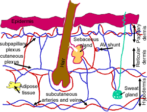
This diagram shows the blood supply of skin.
The circulation of skin
The arteries supplying the skin are deep in the hypdermis. Branches from the arteries pass upwards to form a deep and a superficial plexus.
The deep cutaneous plexus is at the dermal/hypodermal junction. It supplies the fatty tissue of the hypodermis, and the deeper parts of the dermis, including the capillaries for hair follicles, deep sebaceous glands and sweat glands.
The superficial subpapillary plexus lies just beneath the dermal papillae, and supplies the capillaries in the dermal papillae. The pink colour of skin is mainly due to the blood seen in venules of this plexus.
There are many arteriovenous anastomoses in the dermis, which can prevent blood from entering the superficial cutaneous plexus. This strategy is used as a response to cold as a way of conserving heat. The danger is that if the epidermis loses its blood supply for too long, it will die (frostbite!).
Alternatively, when it is hot, more blood is allowed into the superficial plexus, and the skin flushes. The blood in the superficial capillaries is cooled by the evaporation of sweat of the surface of skin.