Microvilli (plural of microvillus)
These are small finger like projections, about 1mm in length, and 90nm or so in diameter. The microvilli are shorter and narrower than cilia.
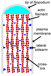
They contain bundles of parallel actin filaments held together into a bundle by cross-linking proteins called villin and fimbrin.
Lateral arms containing myosin I and calmodulin link the actin filament bundle to the plasma membrane.
Microvilli are present on the luminal surface of many epithelia, particularly those specialised for absorption.
Microvilli increase the area of the free surface of the epithelium available for absorption.
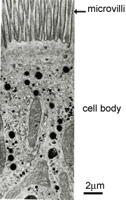
This picture shows a Transmission EM through a tall coumnar cell in the small intestine. At the top of the cell, that projects into the gut lumen, there are the fine structures called microvilli.
Because this is a section through the cell, it does not cut perfectly symmetrically through all the microvilli in the section, and this explains the appearance of the microvilli as shown here. (Think about it!).