Cartilage and Bone: Types of mature bone
Compact Bone
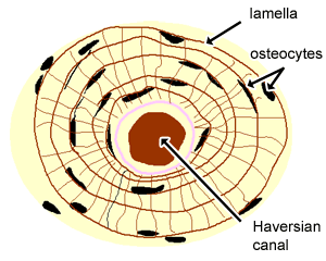
The diagram above shows a transverse view of an osteon (Haversian system) - the basic unit of compact bone.
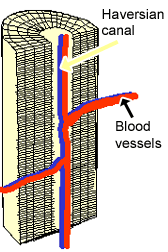
The diagram above shows a longitudinal view of an osteon.
Some, mostly older, compact bone is remodelled to form these Haversian systems (or osteons). The osteocytes sit in their lacunae in concentric rings around a central Haversian canal (which runs longitudinally). The osteocytes are arranged in concentric rings of bone matrix called lamellae (little plates), and their processes run in interconnecting canaliculi. The central Haversian canal, and horizontal canals (perforating/Volkmann’s) canals contain blood vessels and nerves from the periosteum.
This photo shows a cross section through bone. Can you identify the primary and secondary Haversian systems, central canals and bone lamellae?
This is a high power photo of a single Haversian system. Can you identify the concentric lamellae, central canal and the lacunae. Because of the way the bone is prepared for sectioning, you cannot see the osteocytes in the lacunae, only the spaces left behind. The alternating bright and dark concentric rings (lamellae) are due to an alternating arrangement of collagen fibres in the bone matrix. The collagen fibres in each layer are parallel to each other, but at right angles to the fibres in the alternating layers on either side. (Think about how this will affect the bones ability to resist tensile stress).
Cancellous Bone
This photograph shows a section through a marrow space within a bone. Can you identify the bone marrow, trabeculae, osteocytes, and some adipocytes, that are in the bone marrow.
Cancellous bone has large open spaces (marrow spaces) and plates of bone called trabeculae.