Muscle: Cardiac Muscle

diagram of cardiac muscle cells

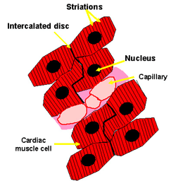
Cardiac muscle is striated, like skeletal muscle, as the actin and myosin are arranged in sarcomeres, just as in skeletal muscle.

However, cardiac muscle is involuntary.

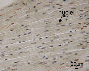
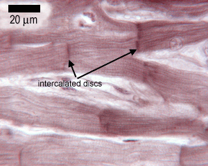
Cardiac muscle cells usually have a single (central) nucleus. The cells are often branched, and are tightly connected by specialised junctions. The region where the ends of the cells are connected to another cell is called an intercalated disc.

The intercalated disc contains gap junctions, adhering junctions and desmosomes.

Gap junctions allow the muscle cells to be electrically coupled, so that they beat in synchrony.

photo of cardiac muscle

This is a low power section through the wall of the heart, showing the striated appearance of the muscle, and the nuclei.

What you can't see very clearly here are the intercalated discs, which are shown in a higher power image below.

photo of intercalated discs

Here, in this higher magnification picture (compare the size of the scale bar with that in the picture above), you can now see individual cardiac muscle cells, that branch. The densely staining regions at the ends of the muscle fibres are intercalated discs.